|
|
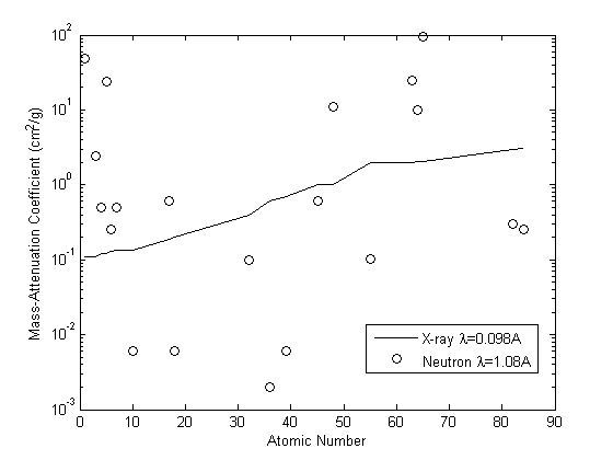
| Fig. 1: Mass-attenuation coefficiences (cm2 g-1) for the elements as a function of atomic number for both X-rays (solid line) and thermal neutrons (circles). [1] |
Neutron radiography is a non-destructive imaging technique that uses thermal neutrons to probe the sample. Unlike X-ray, neutron only interacts with atomic nuclei. Therefore, the attenuation pattern of thermal neutron is different from X-ray. Fig. 1 shows the mass-attenuation coefficient for X-ray and neutron with given wave length. [1] The mass-attenuation coefficient μ/ρ can be defined by Beer-Lambert Law [1]
Here I is the beam intensity at the depth of x in the substance, I0 is the original beam intensity and ρ is the mass density of the substance. From the figure one can notice that the attenuation of X-ray continuously increases with the atomic number, while there is no significant correlation between attenuation of thermal neutron and the atomic number. This feature provides many useful applications of neutron radiography compared to X-ray radiography. For example, the high attenuation of thermal neutron in elements such as hydrogen and carbon will make them shadowed while high atomic mass material such as iron and lead can be penetrated easily by neutron. In the case of X-ray inspection, light element as hydrogen and carbon can be penetrated while heavy metal as lead will be shadowed. Therefore, neutron radiography is widely used in cases like inspection within the shield of heavy metal. Also, the difference in neutron attenuation can be used to identify specific element between neighboring elements that has similar X-ray attenuation. This feature can also be used to identify isotopes. [1]
Most work in neutron radiography is performed with thermal neutron defined as neutron with energy of about 0.025eV. There are two reasons for the choice of thermal neutron. First, neutron within this energy range can exhibit the useful attenuation feature described above; second, thermal neutron can be easily obtained. [1] Neutron from point sources, e.g. nuclear reactor, usually has higher energy than thermal neutron and diverges in direction. Therefore, it is necessary to slow down and collimate the neutron to generate a sharp radiograph with high resolution. Fig. 2 shows a common arrangement of the thermal neutron source. [1]
Many types of reactor can be used as neutron source. For example, H. Berger proposed a reactor using the solution of uranyl nitrate as fuel and a BeO reflector to control the reaction. [2] Accelerator and radioactive neutron sources can be used to build transportable and inexpensive neutron sources for radiography. For example, the 3H(d, n)4He neutron generator is widely used. [3]
To moderate the fast neutron generated from these sources, water, heavy water, oil and graphite are used. Significant gain of thermal neutrons can be achieved by placing uranium immediately around the fast neutron accelerator target, which can result in fast neutron fission. [4]
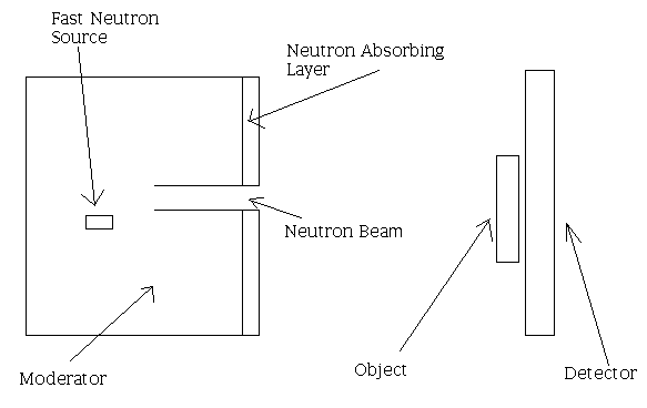 |
| Fig. 2: A method of obtaining a collimated thermal neutron beam from a fast neutron source is shown. It is usually preferable not to have the collimator look directly at the fast neutron source to decrease fast neutron and γ radiation in the beam. |
There are several types of neutron detectors used in neutron radiography. The detector contains converters emitting prompt alpha or beta particles or scintillator emitting visible light. The charged particle can produce a latent image on X-ray or nitrocellulose film. [5] The light emitted by scintillator can be recorded by X-ray film or CCD-camera. [6] Among these techniques, X-ray and nitrocellulose film can achieve the highest spatial resolution. However, this technique suffers from poor contrast and the linearity and reproducibility of the measurements is limited. [7] Also, this technique requires relatively long integration time, which reduces its dynamic resolution. CCD- camera has the highest dynamic range and very good linearity and reproducibility but limited spatial resolution. The dynamic range and reproducibility of CCD can be improved by cooling. [5]
Neutron radiography has become a relatively mature testing technique. Industrial uses can be found in areas involving explosives, plastics and other low atomic number materials contained in metal parts. It is particularly useful in the nuclear industry to identify radioactive isotopes.
© Wenshi Chen. The author grants permission to copy, distribute and display this work in unaltered form, with attribution to the author, for noncommercial purposes only. All other rights, including commercial rights, are reserved to the author.
[1] H. Berger, "Neutron Radiography," Annu. Rev. Nucl. Sci. 21, 335 (1971).
[2] H. Berger, "Characteristics of a Thermal Neutron Beam for Neutron Radiography", Int. J. Appl. Radiation and Isotopes, 21, 407 (1964).
[3] M. R. Hawkesworth and J. Walker, "Review: Radiography with Neutrons", J. Materials Sci., 4, 817 (1969).
[4] A. R. Spowart, "A Mobile Unit for Neutron Radiography", Nuclear Eng., 13, 429 (1968).
[5] H. Pleinert et al., "Design of a New CCD-Camera Neutron Radiography Detector", Nucl. Inst. Meth. Phys. Res. A, 399, 382 (1997).
[6] H. Kobayashi et al., "Neutron Radiography Using Cooled CCD-Camera", in Neutron Radiography 3: Proc. of the 3rd World Conf. on Neutron Radiography, ed. by S. Fujine et al. (Kluwer, 1990), p. 421.
[7] J. C. Domanus, "Research and Standardization Activities of the EURATOM Neutron Radiography Working Group," Risø National Laboratory, RISØ-M-2524, September 1985.